

AFC-330 Non-Mydriatic Auto Fundus Camera
AFC-330 Non-Mydriatic Auto Fundus Camera
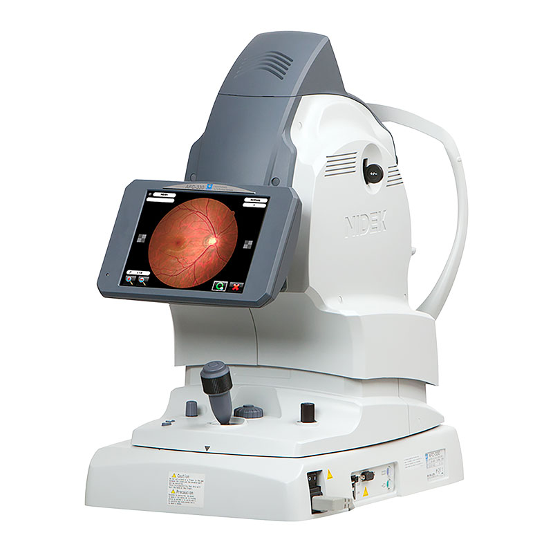
The Marco AFC-330 is a smart fundus camera with built-in camera and computer, automated photography functions, multiple data management utilities, and easy-to-use operator assist functions.
AFC-330 Non-Mydriatic Auto Fundus Camera
The Marco AFC-330 is a smart fundus camera with built-in camera and computer, automated photography functions, multiple data management utilities, and easy-to-use operator assist functions. These smart features make fundus photography easier than ever for screening and diagnosis.
- All-in-one design
- 3D automatic alignment with auto tracking, focus, and capture
- Automatic switching from anterior to posterior focusing
- Automatic panoramic imaging
- Auto pairs for stereo-photography
- External photography
- Low flash intensity and quiet shutter sound
- Auto small pupil mode
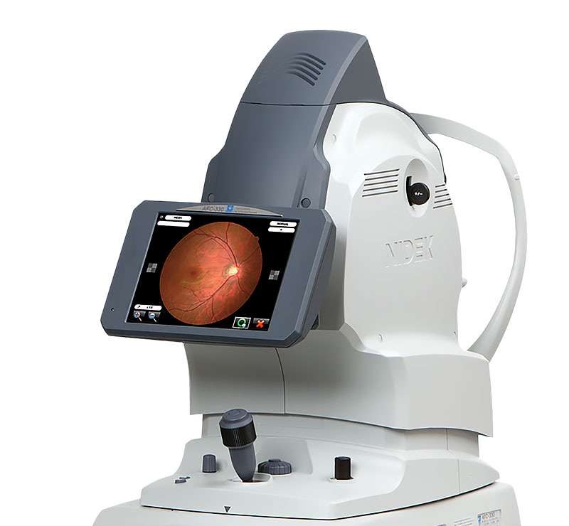
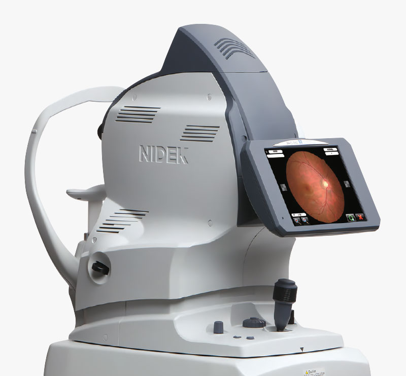
- 8.4” tilting color touchscreen
Imaging Systems /
AFC-330 Fundus Camera
Features:
- All-in-one design
- 3D automatic alignment with auto tracking, focus, and capture
- Automatic switching from anterior to posterior focusing
- Automatic panoramic imaging
- Auto pairs for stereo-photography
- External photography
- Low flash intensity and quiet shutter sound
- Auto small pupil mode
- 8.4” tilting color touchscreen
The AFC-330 Non-Mydriatic Fundus Camera sets the standard in speed, automation, and accuracy.
All-In-One, Space-Saving Device
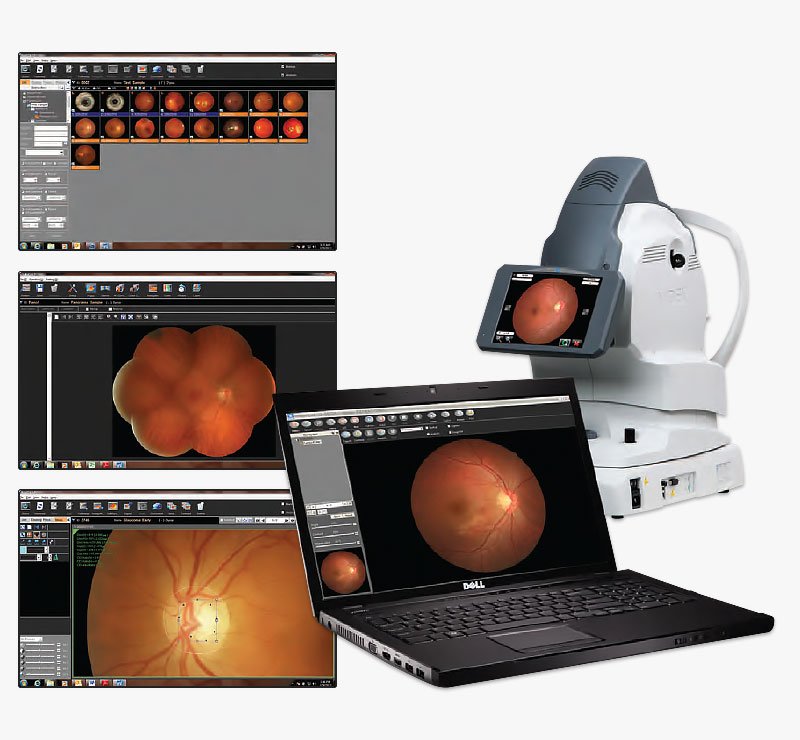
The AFC-330 non-mydriatic retinal camera has an integrated CCD camera and microcomputer in one compact unit without requiring an external camera and PC. This eliminates complicated assembly and wiring during installation. The built-in 12-megapixel CCD camera offers high quality fundus images, and the microcomputer enables easy data management including auto print/export.
Automated Functions for Enhanced Ease-of-Use
- 3D auto tracking
- Auto focus
- Auto switching from anterior eye to fundus
- Auto shot
- Auto print/export
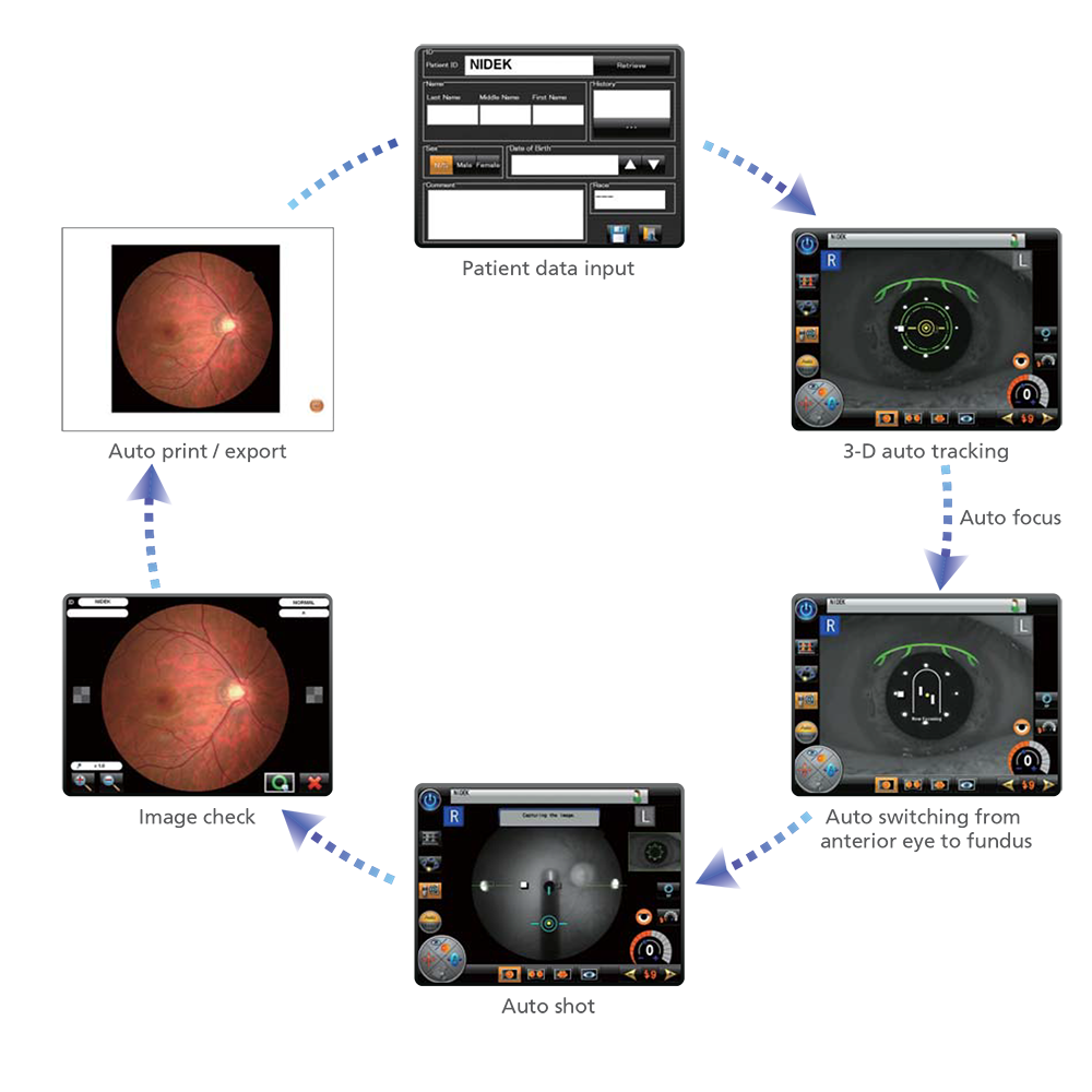
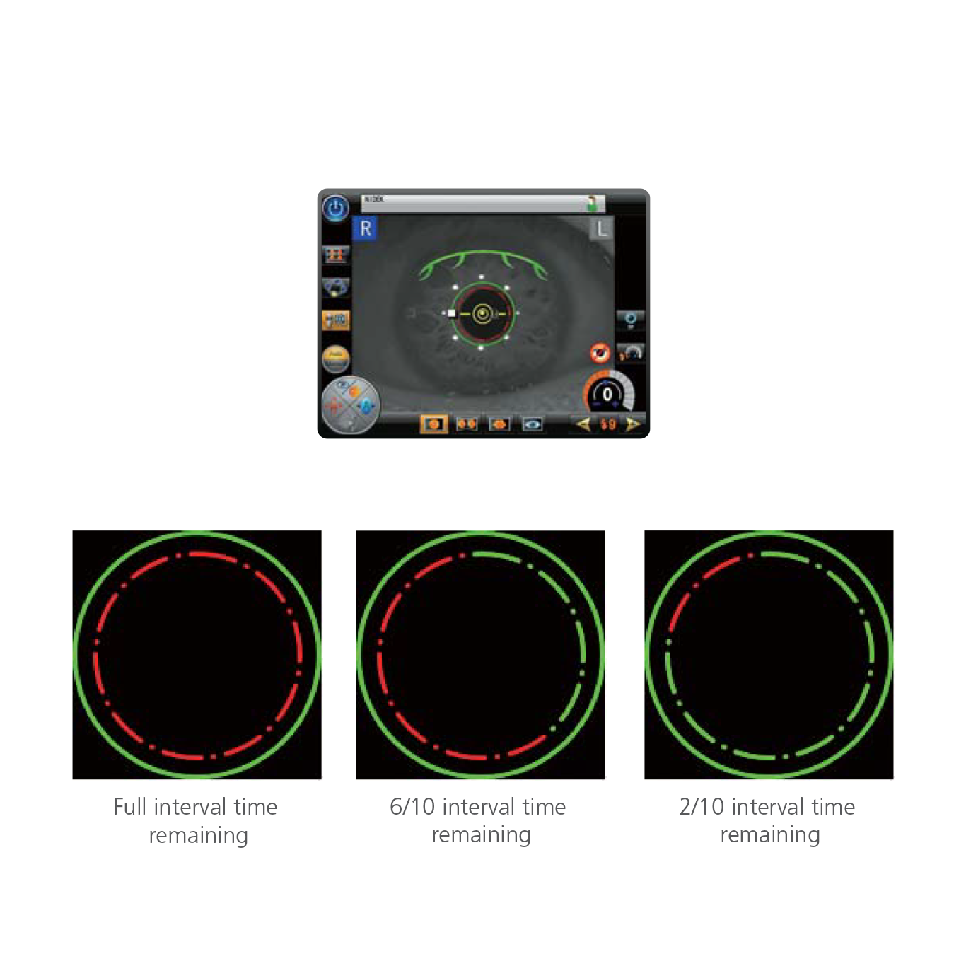
Image Capture Interval Indicator
The image capture interval indicator displays the lapsed time after a shot, which helps an operator wait for an eye to recover from pupil constriction. The interval time can be set from 1 to 10 minutes in 1-minute increments.
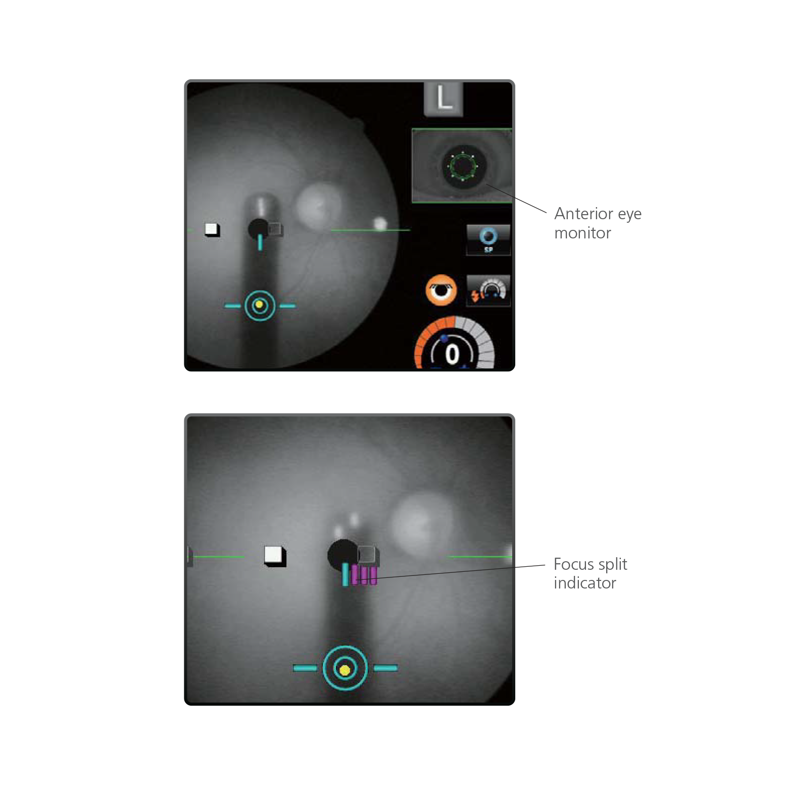
Monitor & Indicator for Operator Assist
The anterior eye monitor inset in the fundus observation screen allows an operator to constantly verify alignment. Additionally, the focus split indicator shows the amount of focus deviation in the fundus observation screen which helps an operator to manually focus the AFC-330 on the fundus.
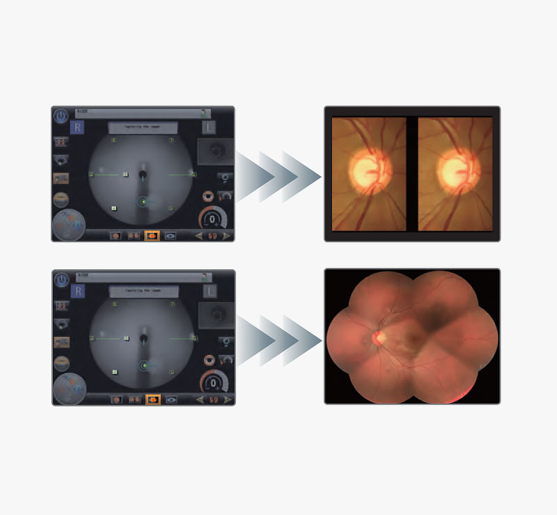
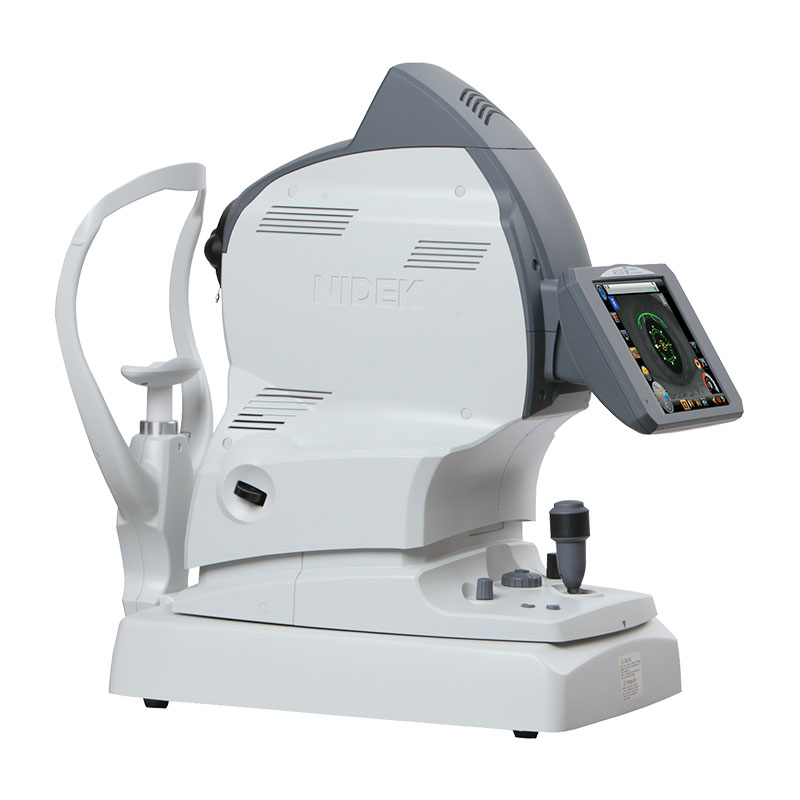
Navigation of Stereo & Panorama Photography
The AFC-330 fundus camera navigates stereo and panorama photography with target marks displayed on the observation screen, which enables an operator to easily capture stereo images and the image series for a panorama composition*.
* Stereo image observation and panorama composition are available with the NAVIS-EX software.
Intuitive Controls
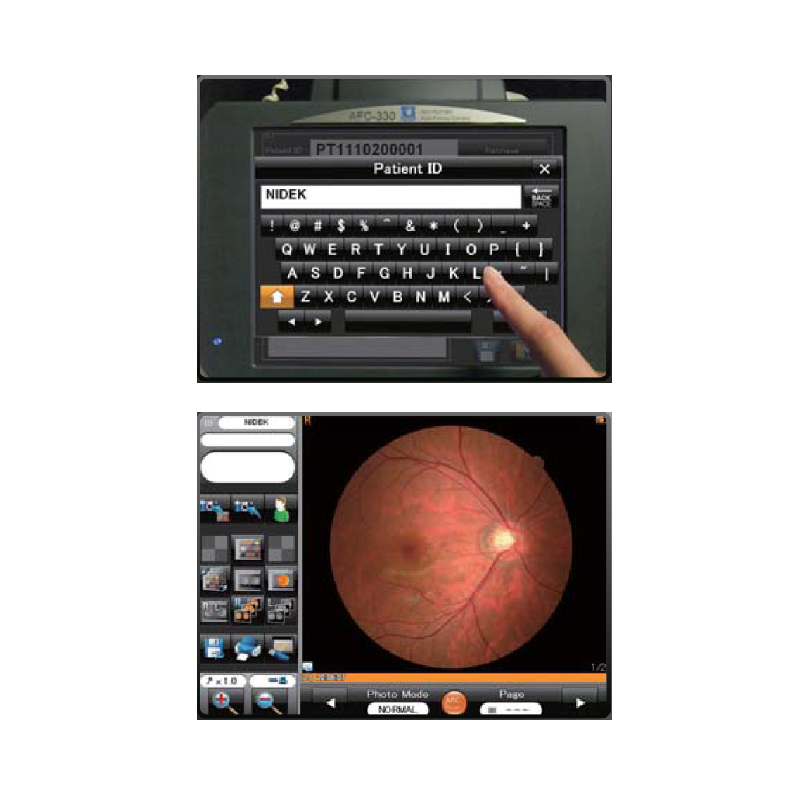
The 8.4-inch color LCD touchscreen display with intuitive menus and icons offers incredible ease of use. An on-screen keyboard enables the operator to input patient data easily without disrupting workflow.
Data Management Flexibility
The AFC-330 offers multiple data management options. Export images across your network using a USB 2.0 storage device and printer, LAN connection with JPEG and XML output, or using NAVIS-EX software.
AFC-330 FUNDUS CAMERA VIDEOS
AFC-330 Non-Mydriatic Auto Fundus Camera Specifications
Technical Details
| Type | Non-mydriatic Automated Fundus Camera |
|---|---|
| Angle of View | 45º (33º in small-pupil photography mode) |
| Working Distance | 45.7mm (from objective lens to cornea) |
| Minimum Pupil Diameter | 4.0mm (3.3mm in small-pupil photography mode) |
| Dioptric Compensation | -33 to +35 D total for patient’s eyes -33 to -7 D with minus dioptric lens -12 to +15 D with no dioptric lens +11 to +35 D with plus dioptric lens |
| Focusing Method | Infrared focus split alignment Adjustable range: -12 to +15 D |
| Light Source | For observation: Halogen lamp 12V 50W For Photography: Xenon flash lamp 300W |
| Flash Intensity | 17 levels from F1 (F4.0 +0.8 EV) to F17 (F16 +0.8 EV) 0.5 EV increments |
| Internal Fixation Target | LED (maximum 9 points) |
| External Fixation Target | Free-arm (optional) |
| Horizontal Movement | 40mm (back and forth) 85mm (left and right) |
| Vertical Movement | 32mm |
| Chinrest Movement | 62mm (up and down, motorized) |
| AutoTrack | X-Y-Z direction |
| Auto Capture | Automatic image capture |
| Camera | Built-in 12 megapixel CCD camera |
| Display | Tiltable 8.4-inch color LCD touchscreen |
| Interface | LAN, USB 2.0 |
Dimensions
| Measurements | 316mm (W) x 518mm (D) x 579mm (H) 12.4” (W) x 20.4” (D) x 22.8” (H) |
|---|---|
| Mass (Approximate) | 29 kg |
Power
| Power Supply | AC 100-240 V ±10%, 50 / 60 Hz |
|---|---|
| Power Consumption | 150 VA |
AFC-330 SUCCESS STORIES


AFC-330 Non-Mydriatic Auto Fundus Camera