Tag: md

Achieve Results and Manage Expectations

It is my belief that an excellent postoperative patient result begins with detailed attention to preoperative testing. With its diverse pre- and postoperative evaluation capabilities, the OPD-Scan III has improved my surgical outcomes and has increased overall patient satisfaction. Furthermore, I use the images on the OPD to discuss diagnoses and treatment plans with patients.

Pre-op Screening for Optimal Post-op Results
To achieve the best postoperative results, we must screen patients preoperatively to identify certain corneal disorders that may affect outcomes. The OPD-Scan III measures a large amount of data, all of which are helpful in determining the best treatment options for patients. We are constantly finding new ways to maximize its advantages. The OPD has led to reduced postoperative issues and complaints by helping me to properly select those patients who would be good candidates for premium IOLs.

For example, the OPD identifies corneal diseases — such as irregular astigmatism, higher-order aberrations (corneal coma or corneal trefoil), high or low spherical aberration, and visually significant pterygiums with irregular astigmatism extending into the central pupillary zone. In each of these situations, the patient would be considered a poor candidate for a multifocal IOL.

I also evaluate the placido rings to look for irregularities. Irregular placido rings can be caused by a wide variety of corneal diseases, including dry eye disease, pterygiums, keratoconus, Salzmann’s nodules, corneal scarring, and epithelial basement membrane disease. If I decide to treat the corneal disease prior to cataract surgery, I obtain a repeat scan after surgery to show patients the subsequent improvement in the placido rings. This helps demonstrate the benefits of having an additional surgery prior to cataract surgery.

The OPD is especially helpful for identifying subtleties of dry eye preoperatively that I may not have otherwise noticed. It is easily seen as irregular placido rings, which can be shown to patients for educational purposes. From there, we begin a more thorough dry eye evaluation. After treating the dry eye disease, I repeat the preoperative testing and IOL calculations. Always address dry eye disease before surgery.

In addition, I evaluate all patients who have had previous LASIK, PRK, or RK. I am looking for irregular astigmatism, high spherical aberration, or a decentered ablation. I discuss these findings with patients preoperatively, explaining that prior refractive surgery may affect their final postoperative result, and because of this, it is possible that not all glare or decreased contrast sensitivity problems will be resolved.

The OPD also measures corneal spherical aberration, which allows me to customize my choice of IOL to best correct it. This helps to optimize each patient’s postoperative visual outcome.

Pre-op Screening of Premium IOL Patients
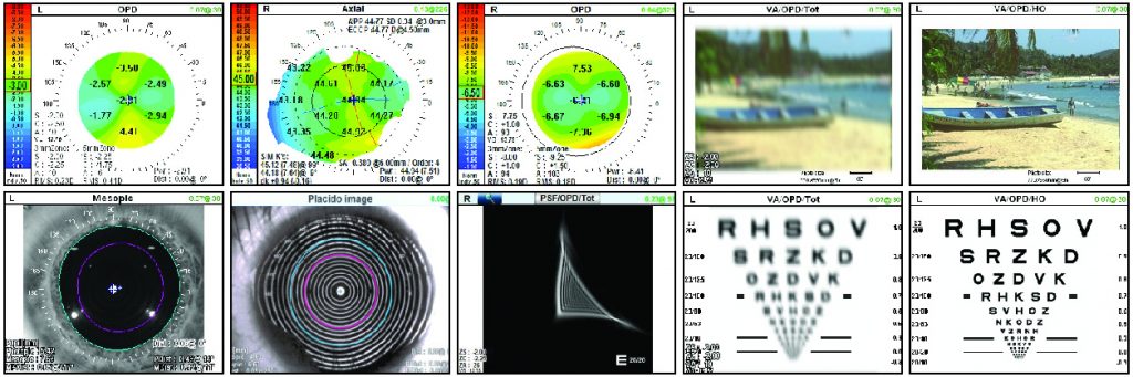
The OPD is especially valuable when evaluating patients who are interested in a toric IOL. The axial map is useful in confirming the planned axis for the orientation of the toric IOL, which should be very similar to the axis obtained via optical biometry. If the axis from the axial map and optical biometry are not similar, I repeat testing and look for the previously mentioned corneal disorders.

The OPD-Scan III also assists me in the multifocal IOL decision-making process. When evaluating multifocal patients preoperatively, I look for corneal disease. In general, the more irregular the cornea, the less likely I would be to use a premium IOL. For example, if a patient has significant higher-order aberrations, I would not use a multifocal IOL. In addition, I evaluate the mesopic and photopic pupil size. If the mesopic pupil size is greater than 6 mm, I have a discussion with the patient about an increased risk for halos and glare postoperatively. However, if the patient has a small photopic pupil less than 3 mm, I generally use a multifocal IOL in which the near vision isn’t dependent on pupil size. I also use the OPD to evaluate angle kappa. If angle kappa is greater than .43 mm, I use a multifocal IOL with a larger central zone.

Consistent Results with Post-op Screening
I obtain postoperative OPD-Scan III studies on all of my multifocal patients. Approximately 1 week after surgery, I obtain a dilated reading. This allows me to see if the central zone of the IOL is centered within the visual axis of the patient. If not, I know the IOL has shifted postoperatively, which frequently results in patient complaints of decreased vision and glare. Complaints usually can be resolved by surgically repositioning the multifocal IOL onto the visual axis.

I will also perform an OPD scan 1 week post-op on patients who received a toric IOL. Again, the scan is obtained with a dilated pupil. In this way, I can measure the axis orientation of the toric IOL to ensure that the toric IOL didn’t rotate away from the intended axis of surgical orientation. Correct toric alignment is critical because the power of the astigmatic correction is reduced 10% for every 3 degrees away from the intended axis of orientation. If there is significant rotation of the toric IOL, I will take the patient back to the OR to realign the toric IOL to the proper orientation.

More Information = Happier Patients
The OPD-Scan III has significantly reduced the number of post-op complaints from patients. With more information to guide me through the entire process, I’ve been able to largely avoid unhappy multifocal IOL patients by identifying the best candidates up front. Before we acquired the OPD, we were inconsistent when making these pre-op decisions.

Making the right decision as a surgeon is only one component of meeting or exceeding patient expectations. It’s also important that I educate patients at each step along the way. Pre-op tests help me manage expectations by educating patients with the aid of easy-to-understand test results. Visual aids help explain variables to improve patient understanding. For example, using OPD results, I can show a patient that his lens is well centered and his toric IOL is well positioned. Patients can see irregular placido rings consistent with dry eye syndrome, which reinforces the need to treat their dry eye prior to surgery.

Imaging can also show patients why irregular astigmatism may prevent them from being premium IOL candidates. With this information and education, patients better understand why you’re making certain decisions, they recognize that you’re trying to make the best decision for them — and they appreciate that.

Customization = Efficiency
The varied capabilities of the OPD allow us to create a specific map for pre- and post-op testing to meet the needs of each patient. We created a customized map to include all the important information we need to preoperatively evaluate the patient. By consolidating all the important measurements on one map, pre-op patient evaluations are much more efficient and allow us to create a personalized treatment plan for each patient. It’s amazing how many options there are with this instrument. We have customized it for our needs, and certainly other offices can adjust their maps to accommodate their needs as well.

We have also developed a comparison map to evaluate pre- and post-op LRI data to evaluate femtosecond laser LRI treatments. The outcome analysis can be used to adjust your LRI nomogram for any under-correction or over-correction of astigmatism. You may also use the comparison map to look for any increase or decrease in higher-order astigmatism, which may occur after an LRI.

Improve Satisfaction and Results
We acquired the OPD-Scan III about 5 years ago in an effort to improve our outcomes and patient satisfaction. It has delivered by helping us to preoperatively detect corneal issues that may affect outcomes, identify the best candidates for premium IOL surgery, improve surgical outcomes, and increase patient satisfaction.

– Dr. Ludwick specializes in cataract surgery and is the Medical Director at Ludwick Eye Center, with locations in Maryland and Pennsylvania. He is also an assistant clinical professor at Penn State Hershey Medical Center

Filed under: Marco BlogTagged with: , ,

Better Information, Better Outcomes

I purchased the original OPD-Scan 8 or 9 years ago, and upgraded to the OPD-Scan III in December 2014. The original OPD-Scan was an absolutely spectacular piece of equipment, and the OPD-Scan III was enough of an improvement that I thought it was worth my while to make the investment. After more than a year, I am confident that I was right.

Better Delivery of Information
The OPD-Scan III is clearly an improvement on what was already great technology. It provides much of the same useful information but in an improved format, and in an easier, more efficient manner. The OPD-Scan III has better resolution and interface, and provides useful, integrated summaries that weren’t readily available in the original model. For example, now my techs can easily obtain a cataract, cornea, diagnostic, or toric summary for me to review and make assessments.

Corneal Wavefront Integration
The ability of the OPD-Scan III to examine the corneal wavefront is a huge improvement, because it helps me to determine the best course of treatment, be it LASIK or cataract. It also helps provide an overview of the patient’s visual status and problems. Every LASIK or cataract evaluation patient has those scans done prior to me even walking into the room, so I have a vast amount of relevant information right in front of me. With better information, I am able to achieve better outcomes for both refractive and cataract patients.

Improved Decision Making
The OPD-Scan III makes my job much easier, and it helps me achieve the best possible outcomes. Let’s say, for example, a patient is in for cataract surgery. I can easily explain to the patient which lens I want to place based on the information that the OPD-Scan III provides. In addition, if I’m considering a multifocal IOL, I can evaluate the corneal wavefront aberrations and quickly make an educated decision as to whether or not the patient is a good candidate for a multifocal IOL, based upon the corneal aberrations that I’m seeing. Or, the information might help me realize the patient is a better candidate for a different lens. I can look at the topography and the regularity of the astigmatism in the corneal surface. The OPD-Scan III is also very good at helping to assess corneal astigmatism. I’m also able to help patients with less-than-perfect vision after cataract or LASIK procedures, whether performed by me or another physician, because data from the OPD-Scan III can help me figure out what is going on with the patient’s vision. It also allows me to see what astigmatism might be coming from inside the eye — either inside the lens and/or the posterior cornea. I have many referrals, complicated patients, or patients who are not seeing well after cataract or refractive surgery, and the ability to take all of these measurements and look at all of the various pieces of information often helps me identify the patient’s problem. This, in turn, helps me make a determination as to what is the right solution for the patient.

Technicians’ Seal of Approval
Because my technicians perform the tests with the OPD-Scan III, it is critical that my staff is proficient in using this technology. It is equally important to me that they like the equipment — and they do. My techs appreciate that the information from the OPD-Scan III is more detailed and the equipment is more user-friendly than the previous model; they have fully embraced its capabilities. And because the OPD-Scan III harvests more than 23 diagnostic metrics in 10 seconds per eye and provides so much integrated information, my technicians often will make a decision for a given patient as to what summary I might need, which is extremely helpful. Sometimes, I have to go back and ask for a different summary or scroll through different images, but that’s simply because there is so much information available at my fingertips if I need it.

An Office Favorite
If you’re looking to provide improved care for your cataract and LASIK patients, there is no question that the OPD-Scan III is worth the investment. The equipment is fantastic, and the customer service that Marco provides is remarkable. The support team comes in to help customize and set up the summaries that will best suit each office’s needs. They are always accessible and helpful. There are many pieces of equipment that I have and love, and I could probably get away with just using them. But I wouldn’t be seeing the whole picture concerning each patients’ optical path. With the OPD-Scan III, I can obtain the basic information — and so much more. There are so many things that this piece of equipment can illuminate for me, that I’m sure I haven’t even scratched the surface yet. It’s my favorite piece of equipment and I can’t imagine practicing without it.

– Dr. Horn specializes in cataract and laser eye surgery at Vision for Life in Nashville, Tenn.

Filed under: Marco BlogTagged with: , ,

Marco’s EPIC-5100: Technology Worth Fighting Over

As a solo, private practice surgeon for the past 25 years, I’ve performed more than 25,000 cataract surgery procedures and more than 30,000 laser vision correction (LASIK, PRK) procedures in my career. After performing some 25 to 30 of these procedures each week, I can tell you that Marco’s EPIC-5100 workstation is an absolute must-have piece of equipment in my practice.

Indeed, we conduct all of our cataract evaluations using the EPIC-5100, which comprises Marco’s OPD-Scan III (a combination autorefractor, topographer, pupillometer, and wavefront aberrometer) as well as Marco’s TRS-5100 digital refractor, autolensometer, and an electronic chart.

Using the powerful EPIC-5100, we can gather all of the information we need, including refractive data, angle alpha, angle kappa, pupillometry (mesopic and photopic), and corneal topography (corneal versus lenticular astigmatism) when considering IOL selection — all of which helps us to make the proper IOL selection for our patients. As a result, we are achieving better outcomes.

We all know that not every patient is a candidate for advanced IOL technology. Patients need to meet certain criteria, including angle alpha/kappa, corneal coma, and pupil sizes. The OPD-Scan III captures the data needed to qualify patients for multifocal, toric, and monofocal IOLs based on their individual visual needs. In addition, the OPD-Scan III gathers all of the information needed to assess aberrations in a patient’s visual system. Dry eye, for example, will induce corneal coma, and being able to show the difference drops can make helps patients better understand this preexisting condition. Pterygia can be monitored and measured over time to know when they are creating aberrations and need to be removed.

Improving Efficiency and Patient Throughput
As an “all-in-one” device, the EPIC-5100 helps improve patient flow and data gathering. Not having to move patients from device to device saves time and provides more accurate data, specifically by reducing the risk of tear film dehydration resulting from the use of multiple diagnostic machines. The EPIC-5100 enables one technician to work up at least 30 patients per day. And with its small footprint, much less space is needed for the EPIC-5100 than an actual exam lane.

We only have one EPIC-5100 workstation because my office space can’t accommodate additional EPIC-5100 workstations. But we plan to purchase two more when we move into our new office next year. We see roughly 50 to 70 patients a day, so two more EPIC-5100 workstations will be incredibly helpful. Other devices also can be added to the EPIC-5100 workstation for further testing, such as for tear osmolarity, increasing its efficiency that much more.

Another advantage of the OPD-Scan III is that it doesn’t require repositioning the patient’s head, as some other products do. When a patient’s head is turned, and he or she is looking at a target, this can induce excyclotorsion, which could lead to erroneous imaging, such as induced astigmatism. The OPD-Scan III measures patients in their natural seated position.

I’m only slightly exaggerating when I tell you the EPIC-5100 station is fought over by all six of my technicians. They will be happy when we add those additional two stations, as this machine truly is the most efficient way to evaluate patients.

Patients Approve
Like our staff, patients enjoy the advanced approach to our workups using the EPIC-5100. In fact, most say they’ve never had such advanced diagnostic testing in past eye exams. Furthermore, our patient education and IOL recommendations are improved because the OPD-Scan III enables us to gather the data so easily, and we are able to show patients their measurements right there in the exam lane using the system’s viewing software. For example, we can highlight areas on the ocular surface where they have dry eye, show them their corneal topography, or display a retro illumination of the eye that shows their cataract. They really get a kick out of that!

Practice Growth
By increasing the number of patients we can see in a day, our practice has realized at least a three-to-five fold increase in profitability, measured in time and patient conversions to surgery with appropriate surgical approaches selected based on our use of the EPIC-5100 technology.

I would be remiss if I failed to mention that Marco has been an excellent company to work with throughout the last 20 years. The representatives are extremely knowledgeable on the technology, and Marco’s training capabilities make the transition and integration quite easy for my technicians — especially when a new staff member joins our practice.

The bottom line: I have found that the EPIC-5100 results in better efficiency, happier technicians, and more advanced decision-making capabilities. Not only does this make for a happier doctor, it has led to better outcomes and happier patients. What’s not to love about that?

– Mitchell A. Jackson, MD, is the founder and CEO of JacksonEye in Lake Villa, Ill.

Filed under: Marco BlogTagged with: , , ,

One Machine… Endless Possibilities

As a veteran ophthalmologist who is still relatively new to running my own private practice, there’s probably not much advice I can offer to my more experienced colleagues. But to my younger peers, I can speak from experience: Go with Marco’s OPD-Scan III as you venture forth to open your own private practice. You’ll be glad you did.

After earning my medical degree from the University of South Florida College of Medicine in 2000, I completed an ophthalmology residency at the University of Florida and my corneal surgery/refractive surgery/uveitis fellowship at the University of California, Davis, where I used the OPD-Scan I and OPD-Scan II for pre-LASIK screening and surgical planning. I also used the two devices for surgical evaluation and planning of corneal-transplant patients. After completing my fellowship, I wenton to practice with a large multispecialty group in Southwest Florida, where I had access to many types of anterior segment diagnostic devices.

In October 2015, I decided to leave that group and start my own practice. To do so, I needed my own accurate and reliable diagnostic equipment. I looked closely at many machines, and test drove several. Ultimately, based on my previous experience with older versions of the OPD and Marco’s unparalleled track record, I selected the OPD-Scan III to accompany me on my new endeavor in private practice.

Fast, Reliable Data
Don’t let the singular name fool you. Like your favorite smartphone, the OPD-Scan III actually consists of five powerful “apps” — an autorefractor, a keratometer, a pupillometer, a corneal topographer with 11,880 data points, and that aforementioned integrated wavefront aberrometer with 2,520 light vector data points.

Moreover, this single device completes 20 diagnostic metrics in less than 10 seconds per eye (including angle kappa and angle alpha, HOAs, average pupil power, RMS value, and point spread function, among others). Wavefront data are gathered from available zones up to a 9.5-mm area, adding the capability to provide for calculation of mesopic refractions. Blue light, 33 ring, placido disc topography is gathered in just 1 second.

Although it’s true that other units in the marketplace can obtain similar images and information,they’re not nearly as simple to use. The OPD-Scan III makes it incredibly easy to create new templates, showing only the information you want to see at any given time for any patient.

As a corneal specialist who actively performs corneal surgeries, such as penetrating keratoplasty and a high percentage of premium IOL and refractive surgeries, I can get all the information I need from the OPD-Scan III for everything from the simplest exams to the most complicated decision-making processes for corneal transplant patients.

With respect to my preoperative cataract surgery patients, for example, I frequently turn to the multifocal IOL (mfIOL) algorithm to assist in IOL selection. This powerful tool provides me with a “decision tree” to confirm in an algorithmic way which specific IOL would be best for a given patient. The algorithm provides me with accurate corneal astigmatism, angle kappa, corneal coma, and pupillary size — all of which play an important role in selecting the proper mfIOL. Indeed, I believe my accuracy and patient satisfaction have increased significantly since I began using this algorithm.

For toric IOL selection, the algorithm gives me another precise measurement that I can compare to other diagnostics, including manual keratometry and my Lenstar biometer. I feel most comfortable when I can compare all of these data, but I would also be comfortable using the OPD-Scan III alone.

Less Space, More Patients
It helps our practice immensely that all of these capabilities fit into an 11″ x 18″ footprint. Since its inception less than 2 years ago, our practice has grown from two employees to seven across two offices in Naples and Fort Myers, FL, and an affiliate office in Miami.

As a relatively new practice in a small office space here in Naples, we have been tasked with seeing patients efficiently and without the luxury of an extra room. The OPD-Scan III allows us to achieve this goal. We see about 100 patients per week at Naples and Fort Myers. In addition, I perform about 30 surgical procedures per week for all three offices. All of the information I need about a patient’s vision can be obtained with this single machine. The unit is definitely space and time efficient.

Impact on Staff
My staff also loves working with the OPD-Scan III. Frankly, they were a little concerned with learning what appeared at the outset to be a complex machine, but after surprisingly short training sessions with Marco representatives, we all were up and running in no time. The same was true with the in-house, hands-on training for our second technician, who required very little ramp-up time (24/7 online training tutorials are also available). For its part, Marco has far exceeded my expectations for customer and technical service. Issues are always resolved quickly. On the few occasions it became necessary, Marco provided a loaner device on short notice, so our workflow wouldn’t be disturbed. Indeed, the loaner backup is a big selling point to me when evaluating other diagnostics and providers.

High Impact on Patients — and Satisfaction
Our patients notice when they sit down in front of one small machine, only to be told a very short time later that we’ve obtained all the information needed for the exam. I can’t tell you how often I’m asked by a surprised patient, “You really got everything you need from just that one exam?” I also believe patient satisfaction comes in the form of successful visual outcomes, especially with premium IOL patients. I know it has helped with our ability to successfully prescribe appropriate IOLs for each patient. Thanks, in no small part, to the OPD-Scan III, we have had a high percentage of premium IOL patients and a very high success rate.

Heed My Advice
As I mentioned at the outset, to those ophthalmologists coming up behind me to start their own practices, I can definitely offer a word to the wise: Go with the OPD-Scan III as you embark on practice ownership. This accurate, state-ofthe-art, all-in-one, time- and space-efficient unit does it all. It eliminates the need for multiple diagnostic machines, and it can be used for corneal and refractive surgeries and evaluations, as well as pre- and post-op planning and evaluations. But don’t just take my word for it — go and see for yourself. You will understand what I mean.

– Dr. Ginsberg is owner and operator of Ginsberg Ophthalmology in Naples, FL.

Filed under: Marco BlogTagged with: , ,